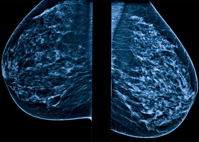
3D mammography is an advanced imaging technique to help discover breast cancer. 3D mammograms provide a clearer view of breast tissues and a better chance of spotting abnormalities than traditional 2D mammograms. With this enhanced detection rate, 3D mammography is an essential tool in safeguarding women’s health.
Why Schedule an Annual Mammogram?
"Routine mammography is a women's single best defense against breast cancer," says Erin Zusan, MD, FACS, breast surgical oncologist. For women with an average risk, the first screening mammogram should begin at age 40, and continue with annual exams. Only 10% of breast cancers are thought to be hereditary, but you should discuss your family history with your doctor to see if they recommend screening earlier.
The survival rate for women with localized breast cancer is nearly 100% thanks to early detection. A yearly mammogram gives you peace of mind knowing that you are staying ahead of your health and that if abnormalities are found, you’ll have a better prognosis.
What to Expect
If it’s your first time having a mammogram, or if it’s been a while since you’ve had one, don’t worry. We do everything possible to make patients feel comfortable, and the process only takes about 15 minutes.
During the mammogram, you will be in a private room with a trained technician. The technician will first perform a health risk assessment by asking you some questions that will help determine your lifetime risk of breast cancer based on the Tyrer-Cuzick model. The questions will cover things like your family history of certain cancers, your medical history, breast density, and information from past genetic testing, if applicable. The results of this risk assessment will be included in your mammogram results.
After the short risk assessment, the technician will help position you for the mammogram. The procedure requires compressing the breasts firmly between a plastic compression paddle and the film. Compression is essential in imaging your breasts to decrease the radiation dosage and obtain clear pictures of the inside breast structures.
Dr. Zusan reassures patients that, “Discomfort during compression is a frequent complaint, but it fades fairly quickly. Our technicians are great and they make the process as quick and comfortable as possible.” During compression, a sensor circles your breast and quickly takes hundreds of X-ray images. This is a safe, low-dose X-ray that can expose changes in breasts that you wouldn’t be able to feel by touch.
Detailed Analysis
A detailed, 3D model of your breast is created from the images taken during compression and then analyzed by a radiologist, a doctor who specializes in imaging. This model allows the doctor to more accurately review and assess any potential abnormalities, especially in denser breast tissue. With this improved precision, doctors feel more confident in their evaluation and are less likely to call you back for a second look.
The images also allow the doctor to determine your breast tissue density, which can only be seen on a mammogram and cannot be determined from a manual exam. There are four main types of breast density: fatty, scattered, heterogeneously dense and extremely dense. Dr. Zusan wants women to understand that, “Knowing your breast density is important because dense tissue can make it harder for the doctor to see abnormalities on your mammogram and can contribute to your lifetime risk of breast cancer.” Women with dense breast tissue may need additional imaging so that the radiologist can get a clearer picture of any changes in breast health.
Understanding the Results
Even if the radiologist didn't see any abnormalities on your mammogram, you will likely see some disclaimers on your after visit summary.
You may see a disclaimer recommending additional genetic testing based on the results of your health risk assessment. Women who have a family history of breast, ovarian or pancreatic cancer may benefit from genetic testing and possibly additional imaging.
You may also see information about your breast density. “We are required to let patients know about their breast density because there are some cases where women with dense tissue could benefit from additional imaging, such as an MRI," explains Dr. Zusan.
"If you see a disclaimer on your after visit summary, it’s nothing to be concerned about. It’s just more informational,” Dr. Zusan reassures. Women can contact their primary care provider or OB/GYN to discuss their results and disclaimers and get personalized guidance on next steps.
Schedule Now or Walk In
If you’re due for your annual mammogram, Community makes it easy to complete this important screening. Visit eCommunity.com/mammogram or MyChart to find a location near you and schedule online today. Walk-in services are also available at select locations.